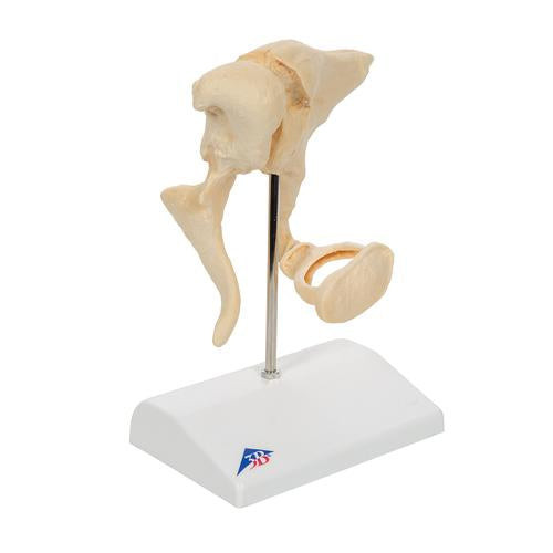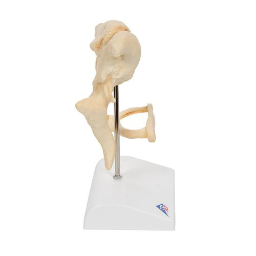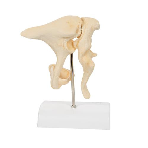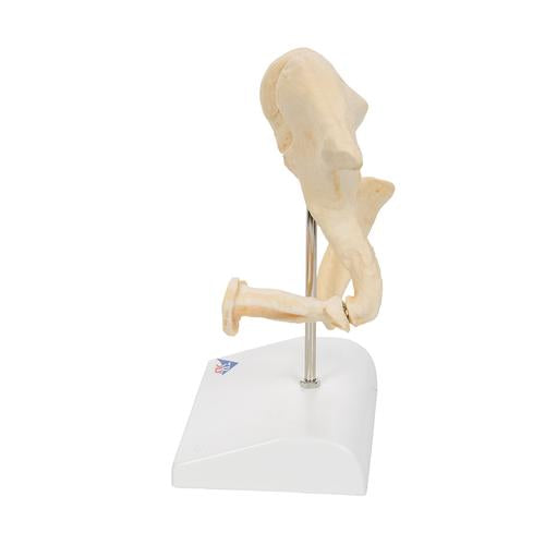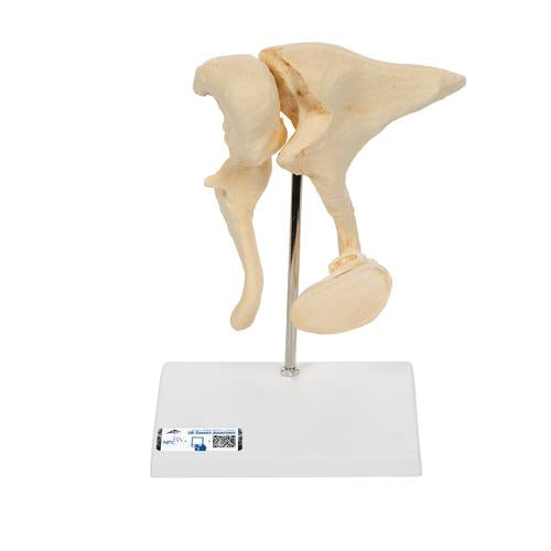
Human Ossicle Model (BONElike), 20-times Magnified - 3B Smart Anatomy
The three smallest bones that are joined to each other in the human body are located in the middle ear and are referred to as the auditory ossicles: malleus (hammer), incus (anvil) und stapes (stirrup). Their job is to transmit incoming sound from the eardrum via the vestibular window to the inner ear and mechanically amplify the sound. The inner ear must be protected from lasting harm which can be caused by loud noise, this is achieved by a reflex that triggers muscle movement (stapedius muscle), causing the stapes to tilt for a short time and therefore sound can only be partially transmitted.
If the auditory ossicles are shaken too vigorously (e.g. a sneeze or a cough), another muscle (tensor timpani) provides protection, by sticking to the hammer and tightening the eardrum. Otosclerosis is a typical condition that affects the auditory ossicles, causing them to stiffen, leading to increasing loss of hearing. In our model, you can see a cast and enlargement of original ossicles, created using micro CT.
Every original 3B Scientific anatomy model now includes these additional FREE features:
- Free access to the anatomy course 3B Smart Anatomy, hosted inside the award-winning Complete Anatomy app by 3D4Medical
- The 3B Smart Anatomy course includes 23 digital anatomy lectures, 117 different virtual anatomy models and 39 anatomy quizzes to test your knowledge
- Bonus: FREE warranty upgrade from 3 to 5 years with every product registration
TIP: You will also receive access to a free 3-day trial to all premium features of the Complete Anatomy app when you sign up for your 3B Smart Anatomy course.
To unlock these benefits, simply scan the label located on your model and register online. All 3B Smart Anatomy features are completely free of charge for you.

