Arthrocentesis Model
Brand: Simulab
SKU ARC-20
Original price
$8,821.40
-
Original price
$8,821.40
Original price
$8,821.40
$8,821.40
-
$8,821.40
Current price
$8,821.40
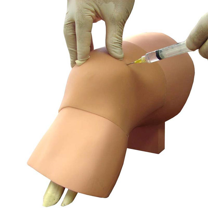
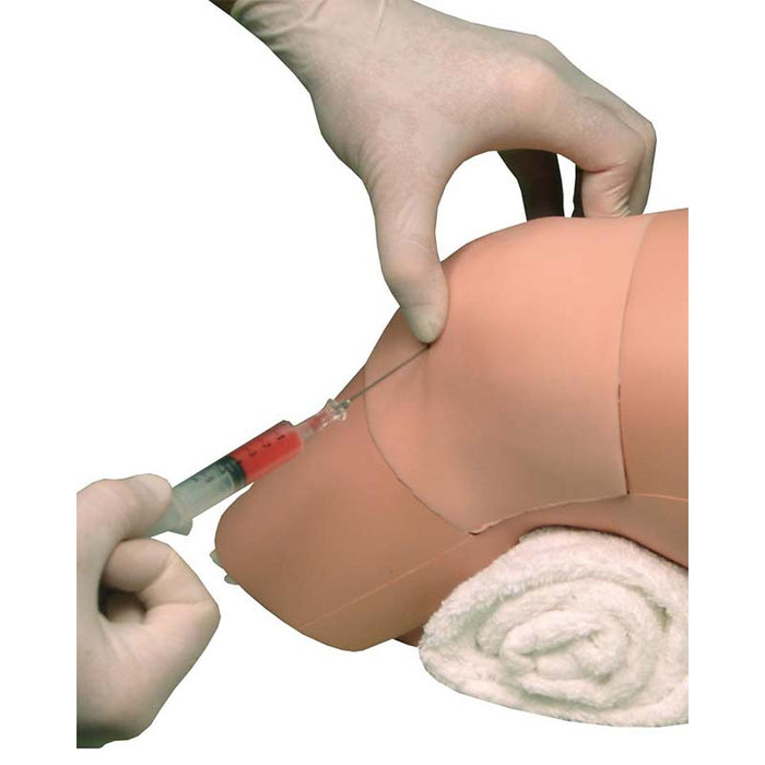
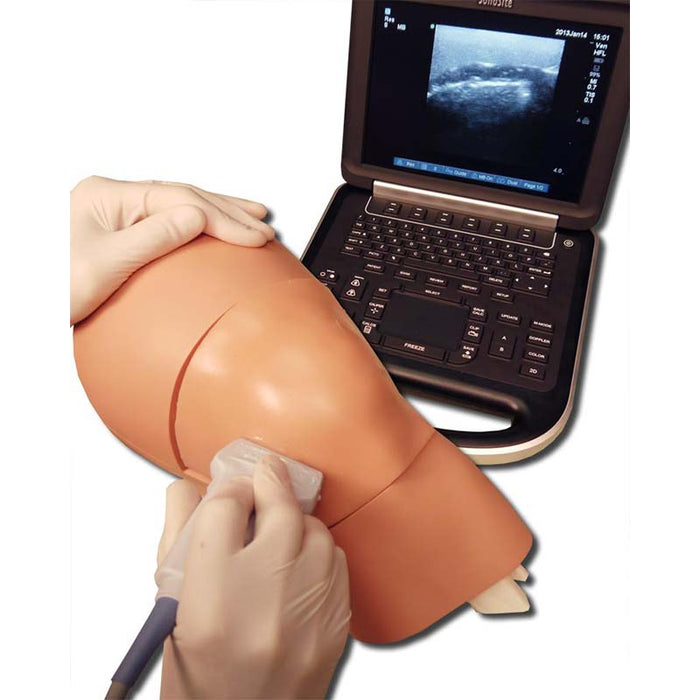
Practice diagnosing and managing the presence of knee effusion with this anatomically correct Arthrocentesis Model.
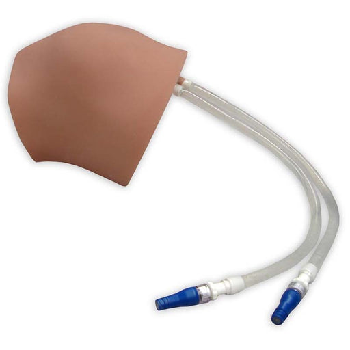
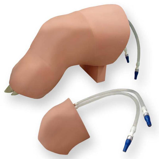
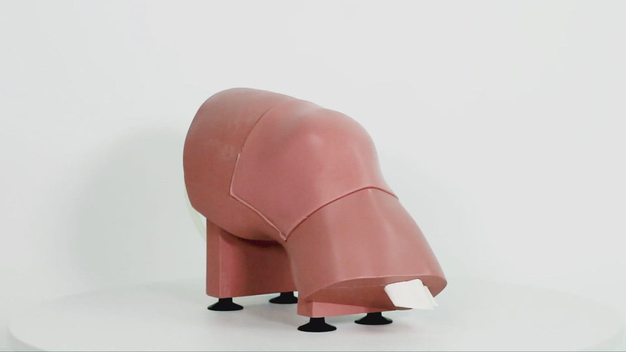
The trainer represents an extended left leg with ultrasound-compatible areas, including the patella, patellar ligament, tibia, fibula, femur, synovial sac, and synovial fluid. Aspirate synovial fluid from a joint cavity using the medial or lateral approach. Insertion sites include suprapatellar and parapatellar access.
Arthrocentesis Model
- Ultrasound compatible
- Highly durable replaceable tissue for multiple uses
- Realistic tactile feedback
- A sensation of bony contact when the needle hits the patella
- A sensation of bony contact when the needle hits the femur
- Inability to aspirate the syringe while the needle tip is in the soft tissue superficial to the joint capsule
- Easy aspiration of joint fluid into the syringe once entry is achieved
- Fluid can be left clear or be colored if desired
- Increase or decrease the size of effusion with up to 60 ccs of fluid
- Learn to ballot or milk the suprapatellar pouch
- Needle aspiration and injection techniques
- Palpating anatomic landmarks significant to the procedure
- Realistic tactile feedback throughout the procedure
Size: 13" h; 8.5" w; 7" d
Weight: 7.5 lbs