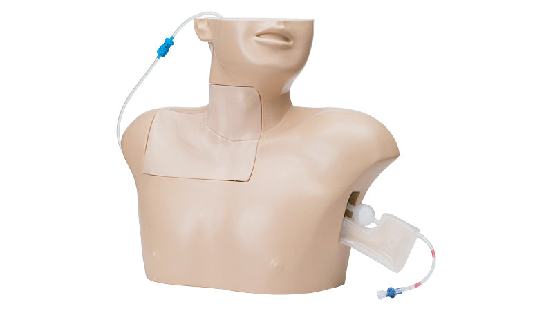
Central Venous Puncture Trainer
Brand: Koken
SKU LM090
Original price
$3,776.50
-
Original price
$3,776.50
Original price
$3,776.50
$3,776.50
-
$3,776.50
Current price
$3,776.50
Designed for practicing ultrasound guided central venous (CV) puncture as well as landmark puncture.
- Enables learners to identify the puncture site by recognizing the important landmarks.
- Simulated blood can be collected when a needle is inserted into the vein.
- Enables learners perform CV puncture by putting negative pressure on the syringe.
- Backflow air pressure indicates incorrect puncture of the lung.
- Since silicone rubber is used for the puncture site, the skin is realistic in external appearance and touch.
- Spare parts are available for various puncture sites.
* This trainer is designed for practicing exploratory puncture. Catheter and guide wire can't be applied.
* Recommed using a needle smaller than 20G.