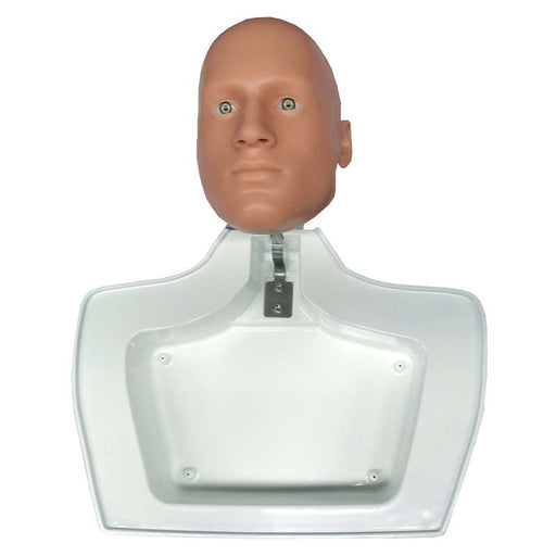
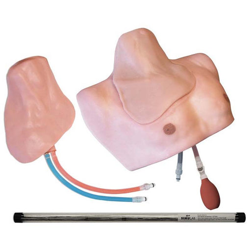
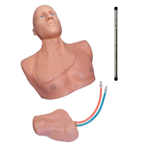
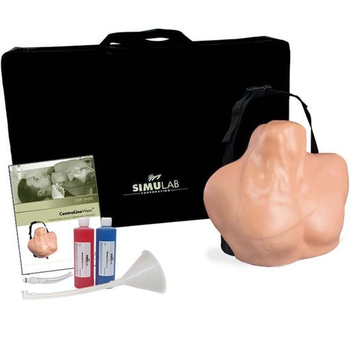
Deluxe Central Line Training Package
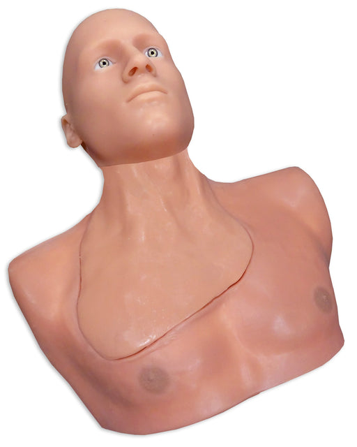
- Anatomically correct, ultrasound compatible tissue, with all relevant landmarks and anatomy.
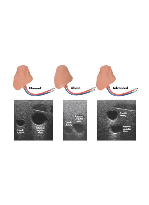
- Enhanced procedural difficulty—Develop user's skills by introducing anatomical variations.
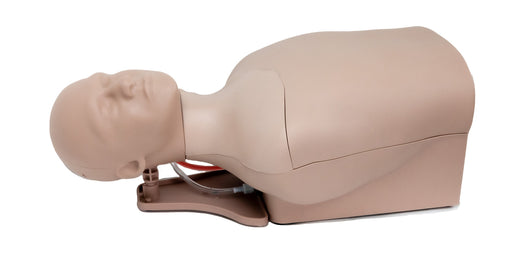
- Enhanced procedural realism—Place simulator in 15° Trendelenburg to practice central line insertion on simulator in standard position.
- Market leading durability—self-sealing tissues and veins offer the greatest value, in the frequency of needle sticks and full catheterizations per access site, of any trainer on the market.
- Exceptional ultrasound imaging through repeated use—needle sticks and full catheterizations do not degrade the image acuity.
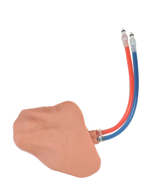
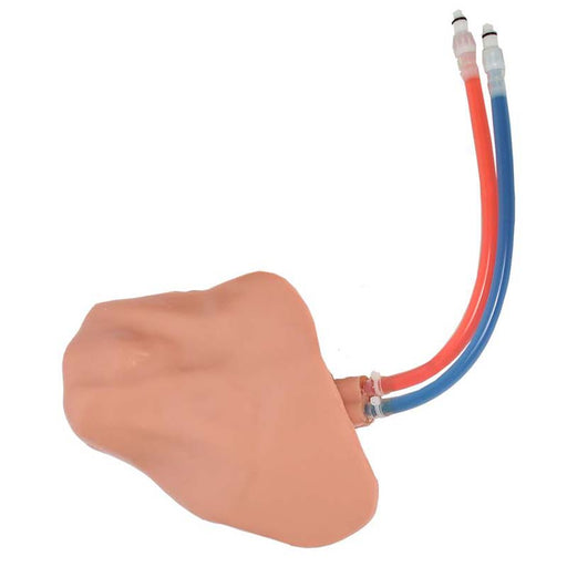
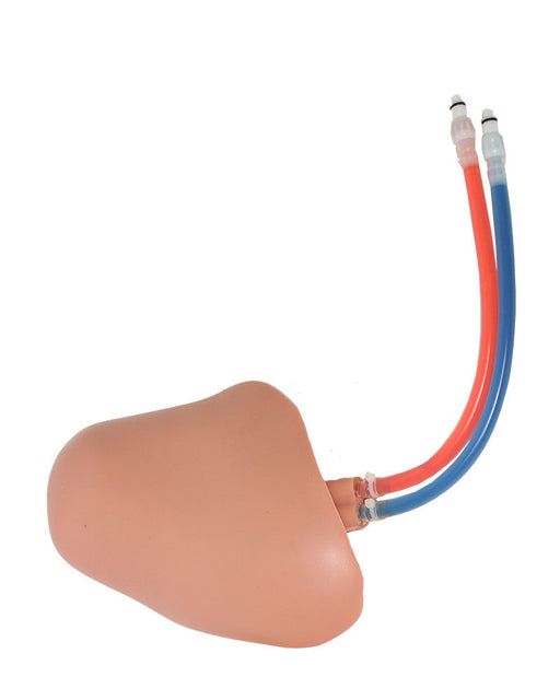
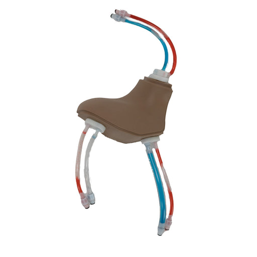
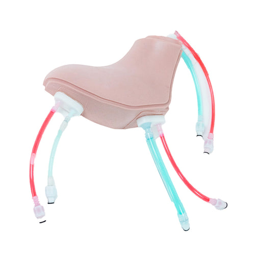
- Two colors of simulated blood differentiate the arterial and venous vessels—provides immediate feedback of unsuccessful cannulation.
- Arterial pulse is present and vein realistically compresses under palpation.
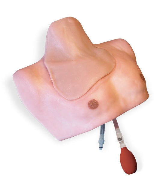
- Easily adjustable venous pressure regulator allows for vein compression or low-pressure simulation scenarios.
- Replaceable tissues come pre-filled with blue venous and red arterial fluid. Tissues can remain filled when not in use, and are easy to refill with provided fluid when necessary.
- Portability—practice simulation in settings of actual patient care.
- Practice full central venous catheterization training—ultrasound-guided or blind/landmark insertion approaches at the subclavian, supraclavicular, and internal jugular access sites on patients with anatomical variations.
- Practice placing the patient in the appropriate position per access site standards
- Gain experience in identifying and selecting appropriate access site based on patient anatomical variations
-
Practice use of ultrasound for:
- Developing psychomotor skills required for obtaining visualization during cannulation
- Detecting anatomical variations
- Distinguishing vessels
- Visualizing arterial pulse and venous compression
- Identifying the anatomical location of the target vessel
- Visualizing needle cannulation of the target vessel in transverse view
- Visualize threading of guidewire in longitudinal axis view
- Visualizing catheter placement
- Reducing the rate of mechanical complications due to anatomical variances such as pneumothorax or arterial puncture.
- Improving first cannulation success and decreasing needle passes
- Practice palpating external landmarks to identify vessel location
- Practice identifying unsuccessful vessel access by fluid feedback representing arterial puncture
-
Demonstrate advanced knowledge and skills needed for insertion on patients with anatomical variations.